Retroperitoneal Hematoma
after
Renal Biopsy
CASE: 52-year-old woman with IgA nephropathy had a renal biopsy
Table (g/dL)
|
After biopsy (days) |
TP |
Alb |
alpha-1 |
alpha-2 |
beta |
gamma |
Hb (g/dL |
CRP |
|
-4 |
6.5 |
3.9 |
0.16 |
0.64 |
0.60 |
1.20 |
11.6 |
- |
|
+8 |
5.9 |
3.2 |
0.24 |
0.59 |
0.83 |
0.96 |
8.5 |
5+ |
|
+15 |
5.9 |
3.2 |
0.25 |
0.45 |
0.86 |
1.10 |
8.5 |
2+ |
|
+22 |
6.3 |
3.6 |
0.20 |
0.50 |
0.78 |
1.20 |
9.4 |
- |
COURSE AFTER THE BIOPSY
The next day of the biopsy, she had a febricula of 37.1KC. The fever was getting higher to the maximum of 38.8KC at the 7th day. Subsequently, although the fever slowly subsided, it persisted until 14th day after the biopsy. An abdominal ultrasonography showed a hemorrhage into the retroperitoneum and a hematoma formation, which must be related with the fever episode.
COMMENTS
In this case, the data of each serum protein, e.g.haptoglobin, was not available. However, the changes in the electrophoretic pattern of the serum clearly showed the process of rise and fall of each serum protein component and the outcome of her hematoma.
Renal biopsy is rather safe in spite of its invasive procedure by which a hemorrhage from the kidney is inevitable. The massive hemorrhage as occurred in this patient was an unusual event. As shown in table, a marked decrease in her hemoglobin of 27% (Hb: 8.5/11.6%) found at 8th (8+) day after the biopsy was equivalent to be no less than 1,000 ml of blood loss from the circulation when her whole blood volume was assumed to be 4,000 ml.
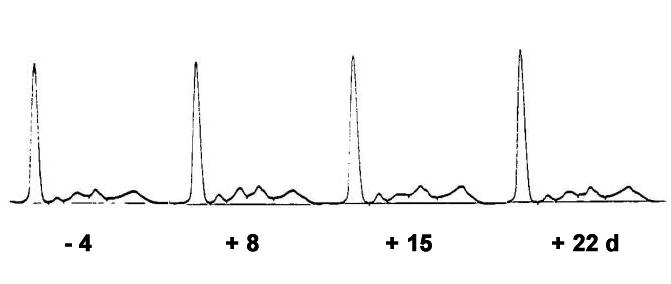
The electrophoretic pattern of her serum at +8 day, about 30 % increases in both alpha-1 and beta globulin fractions, respectively, were found. This must represent the presence of an inflammation due to the tissue damage both in and around the bleeding sites.
At the same time, about 20% decreases found in both albumin and gamma globulin fractions must be the results of loss of her circulating serum protein into the retroperitoneal hematoma.
The change in the alpha-2 globulin fraction of this patient were rather complex. At +8 day, while an about 10% decrease was found in the whole protein concentration of the fraction (Table), a triangle with a sharp summit arose from the fraction (Figure +8). This indicated the following process that, at first, main two proteins of alpha-2 fraction; alpha-2 macroglobulin (alpha-2M) and haptoglobin (Hp); were lost together into the hematoma, and subsequently a rapid synthesis of Hp as an acute phase protein must preceed the not accelerated synthesis of alpha-2M.
On +15 day, the peak of alpha-2 fraction was faded out from electrophoretic alpha-2 fraction, in which Hp must be consumed as the Hp-hemoglobin complex, and the fraction became a low hump mainly composed of alpha-2M.
At +22 day, the electrophoretic pattern of this patient completely normalized, except for the delayed recovery in the hemoglobin. The clinical course shows that it takes, at least, about three weeks to get better from the retroperitoneal massive hemorrhage as caused by a renal biopsy.
REFERENCE
1. Ascenzi , Bocedi A, Visca P, et al. Hemoglobin and heme scavenging. UBMB Life 2005;57:749-59.
T.Inoue Feb 07